You are here
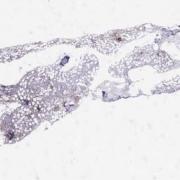
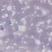
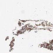
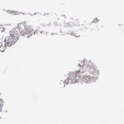
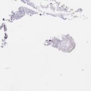
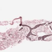
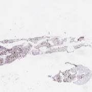
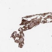
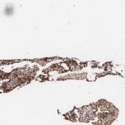
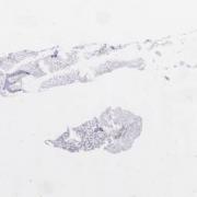
Bone marrow, Acute Undifferentiated Leukemia, CD3 stain [LMP10969]
Clinical History
75-year-old male with pancytopenia.
Clinical Discussion
Flow cytometry showed that the blasts express CD7/11b/38/71/117/123 with a subset positive for CD34. There is no marker expressed here that is lineage specific. Thus, one must consider an acute leukemia of ambiguous lineage. Immunohistochemistry must be performed to rule out other entities of unusual lineage.
Considerations and exclusions in the differential diagnosis:
1) Blastic plasmacytoid dendritic cell neoplasm - excluded due to negativity of CD4 and CD56.
2) AML with miminal differentiation - excluded because there are no markers of myeloid differentiation. We know this entity is negative for myeloperoxidase by definition, however, markers such as CD13 and CD33 should be expressed in this this entity.
3) Acute erythroid leukemia - excluded because the blasts in the biopsy are negative for glycophorin C and hemoglobin A.
4) Acute megakaryoblastic leukemia - excluded because the blasts in the biopsy are negative for Factor VIII and CD61. You should have noticed the interstitial infiltrate of blasts. You must look at the blasts to see if they express these markers not the mature megakaryocytes which would be expected to express these markers. Also, don't overcall the sticky platelets which will also pick up these markers.
Plasma cells were increased but polytypic. The blasts did not express CD20 or CD3. Reticulin shows mild fibrosis (MF1).
Cytogenetics failed on this hemodilute aspirate. NPM1/FLT3 were negative. FISH for chromosome 5 and 7 abnormalities was negative. Only IDH2 mutation was positive but this is not specific enough to assign lineage.
This case illustrates the importance of understanding the markers used in flow cytometry and immunohistochemistry.
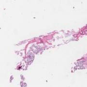
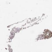
This slide shows CD3 stain of bone marrow biopsy. See related content for peripheral blook, bone marrow aspirate, and H&E and IHC stains of bone marrow biopsies.
Related Content
Wholeslide Image ID:

