You are here
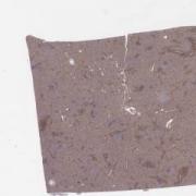
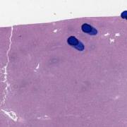
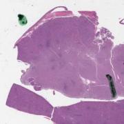
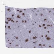
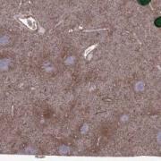
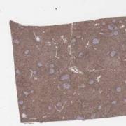
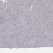
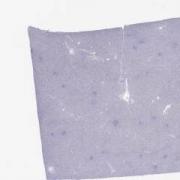
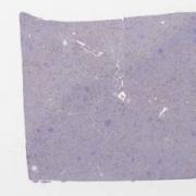
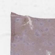
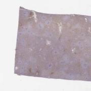
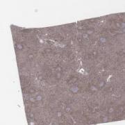
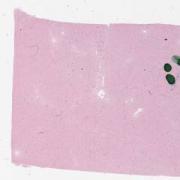
Spleen, Infectious Mononucleosis, CD4 stain [LMP34967]
Keywords
Mono, EBV, atypical, lymphocytes, lymphocytosis, Gammaherpesviral mononucleosis
Clinical History
25 year-old male with splenic rupture.
Molecular studies on the splenic tissue: negative for B-cell clonality, positive for T-cell clonality.
Molecular studies on the splenic tissue: negative for B-cell clonality, positive for T-cell clonality.
Clinical Discussion
Infectious mononucleosis is a benign self-limited lymphoproliferative disorder caused by infection with Epstein-Barr virus (EBV). Symptoms typically include fever, sore throat, lymphadenopathy, splenomegaly, and characteristic atypical lymphocytes in the peripheral blood.
The splenic architecture is preserved but slightly distorted. There is an increase of tingible body macrophages, immunoblasts and plasma cells. Evidence of capsular rupture is also seen. Some immunoblasts may resemble Hodgkin-Reed-Sternberg cells.
Most of the lymphocytes are CD3/CD8+ cytotoxic T-lymphocytes. While the T-cell clonality is positive, the T-cells are reactive. They are not atypical and sometimes, small T-cell clones may be seen in reactive conditions (molecular finding will reveal if oligoclones are present or if a major dominant clone is present, as the latter does not support a benign process). Immunoblasts may be CD30+. EBV is positive in many cells.
The differential diagnosis includes:
Peripheral T-cell lymphoma – but there is no atypia in the T-cells.
Classical Hodgkin lymphoma – but the immunoblasts in this biopsy are not consistent with Hodgkin cells.
The clinical history of splenic rupture also fits with the splenomegaly that can result from infectious mononucleosis. Note that the diagnosis is made after making all considerations. The clinical history is important but should not form the sole basis for the diagnosis.
While the findings are abnormal, the preservation of architecture suggests a benign process.
This slide shows CD4 stain. See related content for other stains.
Related Content
Wholeslide Image ID:

