You are here
Skeleton, Twin reversed arterial profusion sequence [LMP71988]
Keywords
acardia, achrania, monochorionic, reverse profusion
Clinical History
Monochorionic twin of normal fetus.
Clinical Discussion
TRAP is a relatively common (1/100) sequence in monochorionic twins. It is characterized by the presence of one healthy (cardiac) twin, and one severely deformed (acardiac) twin which lacks a functioning heart. The acardiac twin subsists on the cardiac twin’s deoxygenated blood via placental anastomoses between the functioning fetus' arterial system and the recipient fetal venous system. Blood flow is powered in a reverse direction through the acardiac fetus by the functioning twin’s heart.
As such, the acardiac fetus receives only deoxygenated blood, which results in hypoplasia or regression of its major organ systems. Caudal structures are usually more intact in the acardiac twin due to the nature of fetal blood flow. The condition may also result in heart failure in the cardiac twin due to chronic cardiac stress. Surgical intervention may be necessary to save the cardiac twin.
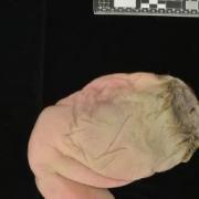
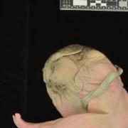
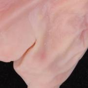
The subject is severely malformed, with no apparent orifices, a single fused lower limb, and a patch of hair above a hyperplastic cranium. Rudimentary rostral-caudal axial morphogenesis is preserved. The mouth is reduced to a small orifice, and the external genitalia are rudimentary. The defect near the "head" is artifact.
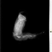
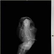
On radiological imaging, subject exhibits an irregular skull, with severe hypoplasia of the calvarium, midface, and jaw. The skull protrudes at an obtuse angle from the spinal column. The spine is highly abnormal, with numerous segmentation defects and irregularly sized and formed vertebral bodies. The sacrum is sharply lordotic. The ribs are highly irregular, with prominent splaying at the costovertebral junction and flaring throughout the ribcage. There are residual hypoplastic radii and ulnae present bilaterally, and aplasia of the humeri, scapulae, clavicles, as well as the bones and tissues of the hands. The pelvis is deformed, with highly irregular iliac wings and aplasia of remaining pelvic elements. The soft tissues of the legs are fused, but show distinct skeletal structures. The femurs have broad metaphyses and are artefactually fractured. The feet are hypoplastic and malformed, with only one completed set of metatarsals and phalanges - corresponding to the first toe - per foot. There is one additional metatarsal corresponding to the second toe bilaterally.
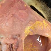
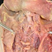
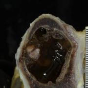
Upon internal examination, two pleural cavities were discovered, with absent lung tissue. There is marked hypoplasia of the gut and urogenital system. The heart and its related structures are characteristically absent. The hypoplastic cranium is fluid filled and largely empty, with some residual brain tissue on the side of the cranial cavity.
This image shows a lateral view radiograph of the fetus. See related content for:
- radiographs: frontal view
- external photographs: front, back, and feet
- internal phtographs: abdominal, pleural, and cranial cavities.
Related Content
Wholeslide Image ID:

